*Note: This is part of a series of thoughts on the topic of looking at movement and movement related symptoms as influenced by the nervous system. These will be dynamic posts with additional content and references being added as time allows, but the primary purpose of the posts are to share my current thoughts on the influence of manual therapy and exercise on what we see and feel in our patients. I hope others will engage me in these thoughts and provide their perspectives and also criticism into the process.

In part 1, I wanted to provide the definition for post-antalgic patterning which I believe is important to understand before thinking about how we treat it (if it even needs to be treated), for which I lay the ground work here:

Post-Antalgic Patterning – Part 2 – A Quick Reference for Manual Therapy and the Nervous System

A little over a year ago Jason Silvernail released a great video summary on manual therapy and the nervous system called “Crossing the Chasm” which definitely had its intended effect on me. This discussion has been a “hot topic” for at least a decade. As I have attempted to share this same information with other clinicians, I have noted a trend towards wanting more “practical” connection between the techniques we use on a daily basis and the nervous system. As a result, over the last year I have started to formulate a way to bring a little bit of clarity to a very complex topic.

Mechanoreceptors – The elephant in the room

In most of our academic preparatory programs for various rehabilitation disciplines, our afferent and efferent sensory nerve fiber education has focused primarily on severe neurological conditions of the peripheral and central nervous system (stroke, spinal cord injury, CNS disease, etc.). However, when it comes to the role of the nervous system in musculoskeletal conditions, the focus tends to be on nocioception (note of importance: nocioceptors are NOT PAIN RECEPTORS!), chemoreceptors (in particular the relationship to inflammatory mediators), proprioception, muscle spindles, and the golgi tendon reflex. We might touch on some afferents when we talk about gate control, but in general, mechanoreceptors are a very minor part of “most” professional academic programming offerings. This is despite that fact that mechanoreceptors may be one of the bodies most densely dispersed points of interaction with our nervous system, in particularly in the tissues we commonly claim to be treating (joint capsules, fascia, ligaments, muscles, etc.).

I remember vaguely talking about Ruffini Endings, Merkel’s discs, Pacinian and Meisners Corpuscles, but I don’t remember much emphasis on them and I certainly didn’t see any value in even recalling their names at the time. Yet now I realize they are probably some of the most important structures I deal with on a daily basis, in particular when it comes to manual therapy interventions. We get so obsessed with the biomechanical properties of soft tissue and joints and the illusion that we can mechanically alter them through our hands and various tools despite growing evidence that this simply is not the case, or at best, has an extremely small role in the big picture. Yet we choose to ignore, or at the very least downplay, the one basic fundamental pathway, the cascade of neurophysiological events which occur every time skin is compressed. These events can result both in short term and long term tissue and movement quality changes which have the potential to explain every single “change” seen through the use of manual therapy. Furthermore, any inflammatory, fluid dynamics, or thermal responses which potentially could come about from an aggressive intervention could have chemical, thermal, and fluid interactions with mechanoreceptors, chemoreceptors, and thermoreceptors thereby compounding and/or altering an existing externally induced neurological stimulus. If the inflammatory, fluid, or thermal process remains active for hours or days, this could yield a sustained stimulus on mechanoreceptors, thermoreceptors, and chemoreceptors thereby influence the nervous system for an extended period of time (think of a “built-in portal e-stim unit” that already exists in all humans).

Perhaps more important than the external stimulus itself is the ability to modify, enhance, and/or guide the therapeutic outcome of the neurophysiologic response from the stimulus with an educational context provided to the patient, allowing for a profound impact on how they perceive touch and movement.

So what does the pathway for this manual therapy to mechanoreceptor stimulus to tissue quality/movement change look like? Dr. Schleip has perhaps best described this in his work on fascial plasticity, of which this diagram provides perhaps the most concise explanation of the relationship between manual therapy and the nervous system.

Schleip, R. (2003). Fascial plasticity–a new neurobiological explanation Part 2.Journal of Bodywork and movement therapies, 7(2), 104-116.

Schleip, R. (2003). Fascial plasticity–a new neurobiological explanation Part 2.Journal of Bodywork and movement therapies, 7(2), 104-116.

To further help solidify the connection between our commonly utilized manual therapy techniques and the nervous system, I put together a couple of acronyms to show the connection between groups of mechanoreceptors and various manual therapy technique:

“RuffMerks need tender care”

  • Ruffini Endings (End Organs) & Merkel’s discs are slow adapting mechanoreceptors which respond best to slow sustained and deep tension
  • ANS (PNS) & CNS interactions
  • General massage, myofascial release techniques, and possibly even ischemic trigger point releases likely preferentially engage these mechanoreceptors

“PacMeisners need action”

  • Pacinian and Meisners Corpuscles are fast adapting mechanoreceptors which respond best to fast & vibratory inputs and are key to texture discrimination (think edged/textured tools)
  • Predominantly CNS interactions although ANS (PNS) possible
  • Greater concentration subcutaneously are also more frequent on the tendinous site
  • IASTM style, cross friction (hand or tool), and oscillating techniques likely preferentially engage these mechanoreceptors
  • Also thought to play a role in high velocity manipulation

 “Free nerve endings do it all”

  • Some free nerve endings are intermediate adapting mechanoreceptors and can respond to any form of touch, or any modality (chemical, thermal, electrical) for that matter.

 “Ligamentous Mechanoreceptors – I got nothing”

  • 4 types, varying adaptability, primarily stretch mediated, although possibly facilitated through touch if the ligament is superficial enough to be compressed
  • Engaged primarily with mobilization/manipulation

Last but not least, how can so called “inert” soft tissue, or fascia, have tissue tension or “tonus”?

Smooth muscle fascia copy

More regarding the existence of smooth muscle cells within fascia can be found here.

To be continued in part 3..

*Note: This is part of a series of thoughts on the topic of looking at movement and movement related symptoms as influenced by the nervous system. These will be dynamic posts with additional content and references being added as time allows, but the primary purpose of the posts are to share my current thoughts on the influence of manual therapy and exercise on what we see and feel in our patients. I hope others will engage me in these thoughts and provide their perspectives and also criticism into the process.

Post-antalgic Patterning – Part 1 – A Definition
Injury occurs either acutely or cumulatively. A threshold is reached and threat is detected, whether conscious or unconscious, the body wants to protect itself. As a part of the physiologic chemical cascade of events which occurs in an attempt to address the potential structural damage, the nervous system, both central and peripheral, protects the region through numerous responses including localized guarding or splinting. This guarding process involves contractile activation of muscle AND the CONTRACTILE activation of what has previously been defined as inert soft tissue, such as fascia, joint capsules, ligaments (1). As a result, kinematics, arthrokinematics, and tissue dynamics may be altered and movement may change. Some of it is subtle, some of it not (2). Regardless, it appears that occasionally, this alarmed state stays active long after the tissue has healed and the threat removed (3).

Steering away from the complicated matter of pain, better discussed in Explain Pain, I wish to focus on what I call “post-antalgic patterning”. This is the existence of an altered movement pattern that is most often an unconscious behavior that remains long after an injury has been healed, or possibly even perceived injury. It exists anywhere in the body with any movement, not just gait! This pattern is a chronic pattern, which begins as early as 2 weeks after an acute injury and remains a minimum of 6 months or for a multitude of years. It may be associated with patient symptoms through regional interdependence, or it may not. Post-antalgic patterning may resolve spontaneously or it may best respond to touch and/or movement coaching. In this definition, any clinician perceived “dysfunction” of the joint or soft tissue contributing to this movement pattern is propagated by the nervous system, not structural change and is minimally influenced by joint or tissue inflammation or swelling.

This pattern may or may not be mechanically inefficient, and it may, or may not, further propagate future episodes of threat elsewhere in the body. In truth, it may just be what it is. Perhaps changing it makes functional improvements and improved symptoms, perhaps not. This is key, because we really don’t understand it, and we have to know when to just ignore it and have the patient move on with life despite this perceived asymmetry, because in reality, we do not know if it might have always been present. This is important, because you have to put limits on how much you try and attempt to alter, as the concept of a “symmetrical” human is fairly illogical. Rather, the objective is to simply to provide an environment to allow movement in a way that the patient can regain trust in these areas, to become more active, which is where the healing occurs.

Your hands or tools aren’t magic. They may or may not be appropriate to providing that supportive environment for altering this pattern, but if you use them, realize their sole purpose is to get the patient moving, reducing threat and letting the tissue re-accommodate to activity.

To be continued in part 2..

1.) Schleip, R., W. Klingler, and F. Lehmann-Horn. “Active fascial contractility: fascia may be able to contract in a smooth muscle-like manner and thereby influence musculoskeletal dynamics.” Medical hypotheses 65.2 (2005): 273-277.

2.) Crosbie, Jack, Toni Green, and Kathryn Refshauge. “Effects of reduced ankle dorsiflexion following lateral ligament sprain on temporal and spatial gait parameters.” Gait & posture 9.3 (1999): 167-172.

3.) Gribble, Phillip A., et al. “The effects of fatigue and chronic ankle instability on dynamic postural control.” Journal of athletic training 39.4 (2004): 321.

This is part 3, the last of a series of posts reflecting on some highlights in learning about movement that I experienced in this last year. In part 1, I addressed my experience with Applied Functional Science / Chain Reaction™ Biomechanics and presented an application of this approach using hip internal rotation. In part 2, I reflected my current thoughts on strength and conditioning. Now in part 3, I discuss my thoughts behind Fascia, Anatomy Trains, and Regional Interdependence.

Much of what we thought we knew about the biomechanical science of fascia and myofascial release is bunk. By saying this, I need to make it clear in advance that this does not change how we treat, rather it changes how we educate our patients and perhaps makes you think more critically about why you might, or might not, want to treat in a certain way. Greg Lehman provides an excellent review of fascial science on his blog.

fascia-man

So what about Anatomy Trains, which I have previously stated may be a beneficial overview for regional interdependence? As Dr. Lehman discussed, it is extremely unlikely that from a manual therapy standpoint we are making biomechanical changes to tissue. If anything, the biomechanical representation of Anatomy Trains better represents fascial adaptation to function and will only respond to progressive overload with daily stresses and exercise. Furthermore, if we look at function and movement, “Form Follows Function” , then the representation presented by Anatomy Trains may vary individually because tissue adapts to the stresses induced on a daily basis.

So we need to throw out the patterns presented by Thomas Meyers, correct? I personally do not think so. This is not the first time we have developed a general map which is not truly accurate of an individual representation. Our good friend the cortical homunculus also is an inaccurate representation of the somatosensory cortex.

Homunculus1

Why? Because the brain is plastic and the somatosensory cortex adapts to how we interact with the world over time, which is most clearly demonstrated by cortical reorganization in phantom limb pain. Yet we still can use the homunculus as a general representation to give us a visual to for understanding sensation. Similarly, I still believe that seeing the patterns in Anatomy Trains can help us better see movement globally and therefore help guide treatment with complex patients representations. From a movement perspective, especially globally, we need to have some way to compartmentalize all the information and how they approximately relate to each other. Joint by joint osteokinematics and arthrokinematics help but can get complex quickly when you combine them with muscles and fascia. Patterns, such as those represented by Anatomy Trains, which encompass both bone and soft tissue, can help compartmentalize and make treatment more efficient if used appropriately. Of course, the reverse is also true, chasing patterns religiously will also take away from the most obvious, efficient, and appropriate treatment approach. Needless to say, these patterns do manifest themselves in our clients and patients from time to time, and to be ignorant of their general representations will cost you and your patient time.

As a side note, we are in a new era of our understanding of pain, with increasing emphasis on a neurophysiological role in this picture. There may be some overlap between the cortical homunculus and fascial adaptation over time. Since fascia is highly innervated with Ruffini and Pacini corpuscles, changes in fascia from physical adaptation to stress and from habituation to particular forms of movement may influence sensory perception and could theoretically be represented in the somatosensory cortex. With some recent evidence regarding the possible existence of a nocioceptive map which overlaps closely with the somatosensory cortex, there is the possibility that sensory rich fascia may be the interface that allows some of our voodoo with regional interdependence to occur, and why sometimes, specificity matters. This is purely hypothetical, but some ground work for the role of fascia and tendon pain (including pain referral) and their related cortical representation  is discussed by Dr. William Gibson (His PhD thesis is available here: Pain sensitivity and referred pain in human tendon, fascia and muscle tissue.) But since we don’t have any other explanation for how manual therapy sometimes requires specificity and sometimes those points of specificity fall in the patterns represented by Anatomy Trains, this is where I am resting my patient education for the time being.

For a better summary than what I wrote above, I highly recommend a post by Alice Sanvito titled:  “If we cannot Stretch Fascia, what are we doing?”.

Sometimes I forget how long the debate over abdominal hollowing (drawing in) vs. bracing has been going on. We’re close to 20 years on this topic. A year and half ago I started writing a clinical commentary on a herniated lumbar disc rehabilitation protocol which included addressing this very topic. A recent resurgence in interest regarding core stability (I guess it never really goes away) made me want to pull an excerpt from the article before it is published. I believe there is a time and a place for abdominal hollowing and abdominal bracing, but there is also a time for neither. The “neither part” has further been bolstered by my experience with Gary Gray’s Applied Functional Science (See Learning about Movement – Part 1). Here is the excerpt:

“Selective recruitment of the TA and multifidi utilizing “abdominal hollowing” or “drawing in” has been described in the literature and is widely practiced. (88,90) In contrast, the simple “abdominal bracing” exercise is another common exercise which not only emphasizes TA and multifidi recruitment, but has demonstrated recruitment of other musculature of the abdominal wall including the internal and external obliques.(89,91) Hodges et al.(85) have demonstrated, using an in vivo porcine model, some increased intervertebral stiffness utilizing TA activation by replicating “hollowing”, more recently, authors have demonstrated that this increased stiffness from “hollowing” is significantly less than during “abdominal bracing” due to decreased activation of the remainder of the abdominal wall musculature. (91,92) When sudden posterior trunk perturbations are introduced, abdominal bracing yielded significantly greater cocontraction of the trunk musculature, increased trunk stability, and better resistance to lumbar displacement than abdominal hollowing.(93) It also appears that the greater the conscious effort utilized to activate the muscular wall, the greater the decrease in spinal stability.(94) Abdominal hollowing has been shown to cause sufficient inhibition of the erector spinae and other musculature to decrease anterior pelvic during hip extension.(95) Inhibition of the erector spinae may have specific therapeutic and treatment benefits in addressing potential muscular imbalances, however they are valuable contributors to stable functional movement, which may result in decreased pelvic control if inhibited.

Although cocontraction of the TA and multifidi has been perceived as vital for muscular stabilization of the lumbar spine, their attempted selective activation may decrease anteroposterior trunk stability,(91-94) theoretically placing rotational trunk stability at risk through decreased activation of the external obliques in comparison to abdominal bracing.(89,91) Despite this, utilizing “hollowing activity” in the early in treatment phases of treatment can provide some symptom relief, kinesthetic awareness, motor control education, and provide some early activation of the TA and multifidi during the acute phase. However, once an individual progresses to postures that involve weight bearing, utilization of bracing may be better to enhance multiplanar stability.

Ultimately, conscious efforts towards bracing and/or hollowing do not contribute toward functional stability, rather, focus should be redirected towards addressing specific motor patterns utilizing multiple muscles in order to develop comprehensive spinal stability.(96) Moreover, from a theoretic functional perspective, consciously constraining movement in any region of the body over an extended period of time may alter the demands placed upon numerous other regions of the body. This may be best illustrated with the influence of external fixation, such as the use of a spinal orthosis for reducing pelvic mobility,(97) single segment spinal fusion influencing segmental motion around it,(98) and the increased risk of degeneration above and below the segment of lumbar fusion.(99) Although these examples entail extreme measures of stabilization beyond the level produced consciously by an individual, it serves as a reminder that a balance between mobility and stability is necessary to enable responses to the dynamic demands during movement of the human body.”

85. Hodges P, Kaigle Holm A, Holm S, et al. Intervertebral stiffness of the spine is increased by evoked contraction of transversus abdominis and the diaphragm: In vivo porcine studies. Spine (Phila Pa 1976). 2003;28(23):2594-2601. doi: 10.1097/01.BRS.0000096676.14323.25.
88. Richardson CA, Jull GA. Muscle control – pain control. what exercises would you prescribe? Manual Therapy. 1995(1):2-10.
89. Richardson CA, Snijders CJ, Hides JA, Damen L, Pas MS, Storm J. The relation between the transversus abdominis muscles, sacroiliac joint mechanics, and low back pain. Spine (Phila Pa 1976). 2002;27(4):399-405.
90. Hides J, Wilson S, Stanton W, et al. An MRI investigation into the function of the transversus abdominis muscle during “drawing-in” of the abdominal wall. Spine (Phila Pa 1976). 2006;31(6):E175-8. doi: 10.1097/01.brs.0000202740.86338.df.
91. Grenier SG, McGill SM. Quantification of lumbar stability by using 2 different abdominal activation strategies. Arch Phys Med Rehabi. 2007;88:54-62.
92. Stanton T, Kawchuk G. The effect of abdominal stabilization contractions on posteroanterior spinal stiffness. Spine (Phila Pa 1976). 2008;33(6):694-701. doi: 10.1097/BRS.0b013e318166e034.
93. Vera-Garcia FJ, Elvira JL, Brown SH, McGill SM. Effects of abdominal stabilization maneuvers on the control of spine motion and stability against sudden trunk perturbations. J Electromyogr Kinesiol. 2007;17(5):556-567. doi: 10.1016/j.jelekin.2006.07.004.
94. Brown SH, Vera-Garcia FJ, McGill SM. Effects of abdominal muscle coactivation on the externally preloaded trunk: Variations in motor control and its effect on spine stability. Spine (Phila Pa 1976). 2006;31(13):E387-93. doi: 10.1097/01.brs.0000220221.57213.25.
95. Oh JS, Cynn HS, Won JH, Kwon OY, Yi CH. Effects of performing an abdominal drawing-in maneuver during prone hip extension exercises on hip and back extensor muscle activity and amount of anterior pelvic tilt. J Orthop Sports Phys Ther. 2007;37(6):320-324.
96. Kavcic N, Grenier S, McGill SM. Determining the stabilizing role of individual torso muscles during rehabilitation exercises. Spine (Phila Pa 1976). 2004;29(11):1254-1265.
97. Konz R, Fatone S, Gard S. Effect of restricted spinal motion on gait. J Rehabil Res Dev. 2006;43(2):161-170.
98. Schwab JS, DiAngelo DJ, Foley KT. Motion compensation associated with single-level cervical fusion: Where does the lost motion go? Spine. 2006;31(21).
99. Putzier M, Hoff E, Tohtz S, Gross C, Perka C, Strube P. Dynamic stabilization adjacent to single-level fusion: Part II. no clinical benefit for asymptomatic, initially degenerated adjacent segments
after 6 years follow-up. Eur Spine J. 2010;19(12):2181-2189. doi: 10.1007/s00586-010-1517-4.

This is part 1 of a series of posts reflecting on some highlights in learning about movement that I experienced in this last year. I hope to be able to do this on an annual basis as a record of self-reflection and hopefully provide some value to others out there on the same journey.

Applied Functional Science / Chain Reaction™ Biomechanics

SKBK_WuShu_Aerial_R

I have previously discussed my interest in going more in depth into multiplanar movement with my posts on 3D stretching as well as regional interdependence. I have used the FMS and SFMA off and on for a few years and felt they were both efficient and useful for their respective purposes, and I love that the research community is actively exploring the validity and reliability of these tests. I will still utilize components of these tests from time to time. However, I have personally found I need to change to a more customized approach to evaluation. In the past, I have found that in the middle of testing, I would break out of the protocols established trying to “tease” out something that did not fit cleanly into any of the tests. Part of this is just my nature, I have a difficult time adhering to standardized procedures and the way I do things just kind of evolves and varies depending by how I perceive something is presented to me. As these breakout sessions grew in complexity, I knew that for me personally I needed to explore some other philosophies which probably have figured out things I have yet to even think to ask about. Gary Gray’s Applied Functional Science (AFS) was the first approach that peaked my interest after that point. Gary Gray arguably pioneered much of the functional training movement, with Gray Cook stating publicly he has been strongly influenced by Gary Gray’s thought process. However, getting to the point of wanting pursue learning about the AFS approach has been a 4 year journey. When I first watched videos of Gary demonstrating and discussing his thought process, I completely disregarded it because my bias at the time of what I was seeing was an awful amount of poor quality movement with no regard for what is currently considered stability, in particular with movement of the spine. But over time, I could not deny that this freedom of motion looked more useful than I had first thought. I finally bit my lip and jumped in, eventually realizing I need to at least give it a try. It was probably one of the best decisions I could have made and now has significantly influenced how I view movement and exercise prescription.

I was given the opportunity to be exposed to the AFS approach through a nine week clinical rotation at Shoreline Sport & Spine in Spring Lake, MI. There are currently 6 Fellows of Applied Functional Science™(FAFS) at this location. A FAFS has completed a 40 week fellowship through Gary Gray’s Applied Functional Science (AFS) approach. The clinicians at Shoreline have integrated AFS with a wide variety of manual therapies and other interventions which was a fantastic eclectic experience that allowed me to explore a number of ways to integrate this philosophy.

Before presenting on this topic, I must first acknowledge that these are my personal reflections on the experience, and if they are in error, they should not reflect upon the excellent clinicians at Shoreline Sport & Spine. I am certain more than one FAFS will perceive I might have missed the boat on key points, and to that, I respond that I plan to formally take a Chain Reaction™ course in the future to see what else I might have missed. Furthermore, I am commenting on but a drop in the ocean of what the AFS system entails. The “Functional Nomenclature” alone requires a 44 page manual to address simply the language and fundamental principles. That being said, here are some key things I learned from this experience:

“Drivers facilitate chain reactions throughout the body”

This was the earliest, most applicable, concept I learned from my exposure to the AFS/Chain Reaction model. It may seem like a simple statement but it is incredibly profound when broken down even a small amount. Rather than simply thinking about one joint moving on another and leaving it at that, the Chain Reaction model demands that every joint be examined from a proximal on distal and distal on proximal perspective, what are the joints above and below doing, and what planes of motion (sagital, frontal, transverse) all of the joints are moving in during any musculoskeletal action. Central to this is the concept that during movement, a driver leads the movement and the joints above and below follow that same movement, but at different speeds as they progressively move in the direction dictated by the driver. This delay in speed/timing of a bone following another bone is the Chain Reaction explanation for much of what we understand about arthrokinematics. When bones move on top of each other in multiple planes of motion in the various representations of roll, glide, spin, etc.,  they are doing so according to the congruencies afforded to them to allow them to follow the next bone and joint as led by the driver.

In most of our manual therapy courses, we examine relative motion of one joint on another and addresses joint movement in various planes of motion based on arthrokinematics. Traditionally, looking at joints this way was left to the manual therapy realm and not the exercise prescription realm. Oddvar Holten likely made the earliest attempt to merge the manual therapy perspective with exercise prescription with his Medical Exercise Therapy (MET – Not to be confused with muscle energy technique) which focused on utilizing various apparatuses to isolate spinal segmental levels and extremities and then focusing on patient induced movement into one or more planes of motion specific to the desired outcome determined in the manual therapy diagnosis. The Chain Reaction approach more broadly addresses this by including concepts more similar to regional interdependence and primarily using the patient’s own body and extremities to control the levels of segmental or joint emphasis through prepositioning such as: Holding on to a stable or unstable support, modifying the weight bearing surface (wedges, angles, instability), conscious prepositioning, etc. It then utilizes another joint or point of the body above or below (could be FAR above or below) to facilitate movement at the joint in the plane, or planes, desired. This approach is both a diagnosis and a treatment, which is the focus of the AFS approach. It integrates extremely well with existing manual therapy interventions, or in Gary’s opinion, independent of traditional manual therapy models, resulting in him developing his own manual therapy system specific to the AFS called Functional Soft Tissue.

Getting back to the idea of a “driver”. A driver is anything which “drives” motor behavior. This could be any part of the upper extremity, lower extremity, trunk, neck, head, eyes, sense organs, and/or even fears and beliefs. The driver itself has numerous variables which can be applied to it: Is it open or closed chain based? What action is performed? What is the direction of movement? What is the speed? What are the force demands? Etc. etc.. This is just a small list amongst many other variables, not even addressing fears and beliefs.  The entire process can get very complex, very quickly, when broken down in the nomenclature which requires looking at every movement from the perspective of:

  • What environment is it occurring in (given available, or specified with certain controls on stability)?
  • What is the beginning position (upright, seated, kneeling, prone, supine, sidelying)?
  • What exactly is the driver (hand, knee, foot, pelvis, trunk, shoulder, etc)?
  • What is the triangulation (direction/target)?
  • What is the action (squat, lunge, reach, pull, etc)?
  • What is the ending position?

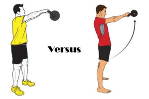
I am not qualified to go into that sort of detail, so instead I will provide a broad overview with a contemporary example. Many of us are already applying general versions of this thought process, but do not realize how far we can take it. I will use the example of a kettlebell swing.

 Kettlebell Swing

If you give a new client a kettlebell and only cue them to swing the kettlebell, they will instinctively “drive” the motion using the arms and shoulders, not their hips as you may have originally intended. They do this because you just gave them a cue which facilitates a motor pattern to accomplish the goal in the simplest way the brain understands, which is to swing their arms to swing the kettlebell, rather than to accomplish the exercise prescription goal you had intended, which was likely hip extension. If you change the cue to “Drive the hips forward”, you changed the driver of the motion to the hips, rather than the arms. Now in order to produce the force to swing the kettlebell, the individual will use a hip extension strategy. You just changed the entirety of the neuromuscular patterns utilized, even though you had the exact same exercise setup. Change the driver and the motor behavior changes.  Now, if you expand this to joint by joint, things get really interesting.

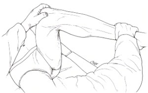
Take for example working mobility and stability of hip internal rotation. There are a handful of non-weight bearing activities which involve the femur actively or passively internally rotating on the pelvis.

ch158f14

We may begin with a manual therapy intervention to address mobility, then provide a mobility exercise, then a stretch, then we prescribe an exercise for stability, then we address another joint which may be associated, and we give it a mobility exercise, and a stretch, and a stabilization exercise, on and on we go. We may end up providing a large amount of exercises, all of which take time, with very specific cues and details to remember. Now with AFS, if we apply the concept of a driver along with any number of subtle changes, or “tweaks” as Gary likes to call them (the process is called “tweakology”), we can tailor a custom exercise specific to our patient needs across multiple joints with reduced need for extensive cuing and details. This can be done with fewer exercises overall because we can integrate mobility, stability, and movement across multiple joints, in multiple planes of motion, into simple exercises which require less time for the patient perform. Progression and regression are simple to teach because you are using movement patterns the client/patient already knows, you simply tweak one or two components to make changes towards the movement you want to improve.

As I am already far over my target word count for this post, I will finish with a video in which I discuss some basic strategies to emphasize hip internal rotation in weight bearing and function:

[youtube http://www.youtube.com/watch?v=wvHEGrUs68o&w=420&h=315]

A few months ago I talked about the Gluteus Maximus Activation Enigma and the conflicting information obtained on the glute max in the clinic versus what has been demonstrated in literature. It has been difficult for me to address this because I too was guilty of really perpetuating the idea of “gluteal inhibition” and that your “glutes are shut off”, when the evidence for these theories does not exist unless you have a true nerve lesion. It may seem like semantics to the some, but the reality is that our patients and clients take these words very seriously. In fact, I would say a good chunk of them catastrophize the fact that their “glutes aren’t working” and likely worsen the associated symptoms involved in the hip extension dysfunction. I think for athletes in particular to be told that something isn’t working in their body is detrimental to performance for individuals with certain psyches, a point which Vern Gambetta really drives home with his opinion on corrective exercise. At the same time, even if the glutes truly are not “Turned off” or “Firing in the wrong order”, clinically, they are clearly not working very efficiently either, especially if they are significantly asymmetrical. Therefore to find middle ground, I like to look for solutions which help the client/patient remain independent while still participating in their sport even if some form of dysfunction exists by using self evaluation and treatment. I previously mentioned my suspicion that muscle fatigue, rather than muscle inhibition or activation order, may play a part in why our glute emphasized treatments result in reduction of symptoms. A recent article from Hong-You Ge, et al.1 demonstrated that latent trigger points have measurable effects on muscle fatigue made me want to revisit fatigue in the evaluation and treatment of general hip extension dysfunction.  However, I’m going to broaden this idea even further (I’m once again breaking my own rules regarding excessive extrapolation of a research study by doing so) by first looking at addressing the antagonists to hip extension, the hip flexors, prior to attempting to address trigger points/restriction in the gluteals.

I want to preface this write-up to make it clear that I have no evidence for the process that I am about to describe and I am certain there are at least 10 other ways to independently evaluate hip extension. I think both Stuart McGill and Bret Contreras have touched on the use of  different types of bridges in determining hip extension dysfunction in the past, but I couldn’t find the articles offhand, so here is my take on it.

I use a 15-20 rep range of single leg bridges for the patient/client to subjectively identify whether they feel a perceived difference between sides relative to fatigue, ease, and whether it feels disproportionately loaded on the hamstrings, possibly even painful if that is their primary complaint. Then, based on which side is perceived as more challenging, we slightly butcher the classic Janda lower cross syndrome2 and just associate hip flexor involvement with gluteal function rather than look at his original cross of abs to glutes.We’ll generalize it even more and call the hip flexors over active antagonists with possible active or latent trigger points in them decreasing performance of the agonist hip extensors just to integrate the Hong-You Ge et al. 1 discussion a little more.

So for the patient to independently treat this, we start with them attempting to inhibit the hip flexors through a 30 second static stretch for and then retest the bridges. They don’t have to go all the way to 20 reps but they should just be able to go 2-3 more reps more and perceive the exercise as easier. If it does improve, have them do a full minute of static stretching of that hip flexor followed by 3-4 sets of 15-20 reps of single leg bridges to reinforce the more efficient hip extension pattern.  If it doesn’t improve, or they feel only a little better, try a self-TFL release next. Use 1-2 minutes of self release on a tennis ball followed by the same 3-4 sets of single leg bridges discussed earlier.  If they still don’t feel an improvement, go for the glutes directly with a self release. If it works, follow the same pattern of reinforcement from earlier. If there is no change, there is a slim possibility they simply need to train that side more aggressively in hip extension. If this is the case, then we want to have them work on quality reps of single leg bridging on a daily basis for the same pattern of reinforcement as described above. If within one week of working this pattern they still find a single set is fatiguing, the problem does not lie specifically in the hip musculature and it is going to require a bigger picture perspective and likely more involved manual therapy (starting with a pelvic/lumbar eval).

[youtube http://www.youtube.com/watch?v=cPebQCkSLCs&w=420&h=315]

A couple of notes: First off, verify that the fatigue is not just related to the position of their foot and whether they are driving from the heel versus the toes because this can significantly impact loading of the hamstrings between sides.  Second, I recognize not every one of our clients and patients can even do a single leg bridge, let alone 20 of them, but this test and these self-treatment options is not for those individuals anyway. Third, by the 3rd set of bridges, if they’re not used to doing these bridges, they’re going to be fatigued anyway, just do a couple reps for them to subjectively evaluate any chance in the performance of hip extension.

Finally, I admit I am probably still going to use the terms gluteal inhibition from time to time, but I swear I’ll do my best to not give patients or clients the anecdote that their glutes are “shut off” again.

***Update 6/24/12: A great example of when self treatment for hip extension dysfunction fails and more involved manual therapy is needed  from Bill Hartman is found here on his blog.

1. Ge H, Arendt-Nielsen L, Madeleine P. Accelerated muscle fatigability of latent myofascial trigger points in humans. Pain Medicine. 2012:no-no. doi: 10.1111/j.1526-4637.2012.01416.x.

2. Janda V. Muscle strength in relation to muscle length, pain, and muscle imbalance. International Perspectives in Physical. 1993:83-97.

The FDA dictates specific uses and indications of a drug based on pharmaceutical trials. These specific uses and indications are “on the label”. Physicians frequently utilize pharmaceuticals “off label” from their branded purpose based on clinical reasoning. This is a legal and widely accepted practice and necessary to treat conditions which at this time may not have the research evidence available to support the practice but has demonstrated good clinical outcomes. The advent of Clinical Prediction Rules (CPR) and subclassifying of conditions towards specific treatment protocols has been growing in the physical therapy realm5,8,12,13. This is particularly true with manual therapy and CPR for joint mobilizations/manipulations for the cervical, thoracic, and lumbar spine. In general, these guidelines tend to be region specific, IE: a lumbar manipulation for a lumbar condition5,8and cervical spine mobilization/manipulation for neck pain12. Although not as well known, predictive factors for the influence of cervicothoracic manipulation on shoulder pain10 and lumbopelvic manipulation in patellofemoral pain syndrome9 have also been proposed. You could say that the advent of CPRs/classifications is the rehabilitation world’s attempt at providing an “on label” guideline for treatment. However, little other attempt has been made to provide subclassifications for conditions and treatments regarding manual therapy interventions on extremity conditions. Clinically, worldwide, many movement professionals treat extremity conditions one to two joints proximally or distally, in particular through addressing mobility at the spine. This practice is based in the idea of regional interdependence, or “the concept that seemingly unrelated impairments in a remote anatomical region may contribute to, or be associated with, the patient’s primary complaint.”18 In an essence, this practice is “off label”, but unlike the pharmaceutical practice, it is not widely accepted and frequently questioned. This is particularly true from a medical billing and, depending on the location, referral/medical prescription level. Even from within our profession itself, it is not terribly uncommon for the concept of regional interdependence to questioned and perceived as a “wild goose chase around the body” 18.  As I have mentioned in previous posts, Thomas Myer’s Anatomy Trains based system KMI, Gray Cook’s SFMA and Gary Gray’s Chain Reaction Biomechanics™/GIFT Fellowship are perhaps the first to develop standardized evaluation and treatment methods of looking at the body globally rather than locally. Although the terminology varies between each, all of these programs have essentially provided a road map towards understanding regional interdependence. I have minimal exposure to all of these programs, so I cannot give justice to any of them trying to give additional details from their models. However, I wanted to share my thoughts on regional interdependence based on the experience I have gained through my mentors, research, and my limited full-time clinical experience thus far.

A JOSPT guest editorial by Wainner, Whitmann, Cleland, and Flynn titled Regional Interdependence: A Musculoskeletal Examination Model Whose Time Has Come (Freely available directly from JOSPT) written in 2007 probably first popularized the term “Regional Interdependence”, because very little literature prior to this date utilized this term. This editorial presented a great case, both from a clinical and a research perspective, that the practice of examining musculoskeletal conditions beyond the single joint/primarily complaint area is woeful inadequate to address the needs of both common and complicated conditions 18. Research has increasingly been supportive of regional interdependence. Improved pain scores and functional outcomes have been demonstrated painful shoulder conditions as a result of the use of cervicothoracic and rib manipulation 2,4,10,15. Similarly, lumbar and pelvic manipulation has demonstrated improvement in patellofemoral pain syndrome9,16,19. Beyond manual therapy, evidence for the use of foot orthotics for various lower extremity injury as a preventative measure7 and as a method of treating PFPS 17.  However, the role of distal contributions, or more specifically excessive pronation, was recently questioned in a systematic review by Chuter and Janse de Jonge6. In their review, they proposed that a greater influence on lower extremity injury arises proximally from the “core”6. Regardless, what is evident in available research is that proximal and distance regions to the site of injury have some role either as a result of the injury, or as a precursor to the injury1,6,11,14.

Clinically, there are presentations and treatments related to regional interdependence which are a long way (if ever) from being able to be demonstrated or clearly explained in a research design. We are still in the early stages of understanding manual therapy, let alone regional interdependence. Bialosky, et al. (Open access link) provides a great review and proposed a model which encompasses both joint and soft tissue mobilization/manipulation 3.  Although the emphasis of this model and much of the research on manual therapy is based on a neurophysiological, peripheral, spinal, or supraspinal mediated mechanism 3, it is difficult to extrapolate whether these the neurological models also play a key role in regional interdependence. Perhaps now, with the treated “dysfunction” one or more joints away from the injury location, the importance of “movement”, as biomechanically dictated, plays a more important role? Or perhaps still, somewhere in “homunculus land”, a map of regional interdependence is now changed to alter both pain and movement patterns. It is too early to tell, but hopefully this question will soon answered! Whatever the mechanism may be, clinically, there still appears to be a degree of specificity and clinical reasoning necessary in order to provide an optimal outcome. To illustrate this, I want to present a brief clinical case.

This case involves a 23 year old male competitive soccer player who originally presented with posterior left rib pain around T5 region which somehow evolved into some form of left posterior shoulder pain and restricted ROM. Somatic dysfunctions for the thoracic spine, ribs, scapula, glenohumeral capsule, and surrounding tissue(including an incredibly tight latissimus dorsi) were identified. These factors were assumed to be key to recovering the 10-15 degree loss of shoulder flexion with a painful posterior “pinch” at the endrange. A gambit of joint mobilizations and attempts to lengthen the latissimus dorsi, as well as, various techniques for “releasing” other soft tissue restriction was started.  Despite 4 sessions of valiant attempts to regain this loss of shoulder flexion through manual therapy and stretching even up to two joints away, little progress was made. Out of shear randomness, I observed an obscurity in the way the inferior aspect of his rib cage moved when I passively flexed his left shoulder. Perhaps it was an illusion generated by my mind from years of staring at Thomas Meyer’s Anatomy Trains, but something made me believe it was worth looking at his rectus abdominis. Needless to say, simply palpating the rectus abdominis was enough to generate a startle response similar to a typical trigger point presentation. Tension and “restriction” was felt through the lateral band of the rectus abdominis. Much to the patient’s dismay, I spent two minutes “releasing” this restriction and without any other treatment. Immediately afterward, I was able to move him into those last 10-15 degrees of shoulder flexion pain free. One additional treatment was scheduled and the patient was set for a one week recheck, at which point they were still symptom free and discharged.

Looking back now, I could pretend I know what happened and propose a theory to explain it from a biomechanical model utilizing Anatomy trains. I could state that since the rectus abdominis inserts on ribs 5-7, it must then pull on the fascial origins of the pectoralis minor directly or through pulling the rib cage down. Consequently, the pec minor then pulls on the coracoid process of the scapula, which could  result in anterior tilting of the scapula, and therefore give a possible mechanical cause for the “pinching sensation” and  restricting shoulder flexion. The honest truth is, I don’t know why it worked, because it was such a random find. Yet oddly enough, it seemed as though I had to be specific enough in my treatment approach in order to get a positive outcome for this patient. Simply addressing classic restrictions around the shoulder was not enough in this case, I had to go even farther, and I had to use soft tissue! Did I truly decrease the tension in the rectus abdominis and therefore produce the mechanical cascade which lead to this resolution? Could it have been placebo, was the shear randomness of the treatment approach a psychological effect that somehow modulated the pain or ROM changes? I am completely open to any suggestions!

Clearly not every case needs to be this involved or complicated, and sometimes the area of injury is the best place to focus your treatment and leave it at that. At the same time, both clinical and research evidence seems to be paving way the importance of remembering that the leg bone is connected to the knee bone, and the knee bone connected to the thigh bone…

1. Berglund KM, Persson BH, Denison E. Prevalence of pain and dysfunction in the cervical and thoracic spine in persons with and without lateral elbow pain. Man Ther. 2008;13(4):295-299. doi: 10.1016/j.math.2007.01.015.

2. Bergman GJ, Winters JC, van der Heijden GJ, Postema K, Meyboom-de Jong B. Groningen manipulation study. the effect of manipulation of the structures of the shoulder girdle as additional treatment for symptom relief and for prevention of chronicity or recurrence of shoulder symptoms. design of a randomized controlled trial within a comprehensive prognostic cohort study. J Manipulative Physiol Ther. 2002;25(9):543-549. doi: 10.1067/mmt.2002.128373.

3. Bialosky JE, Bishop MD, Price DD, Robinson ME, George SZ. The mechanisms of manual therapy in the treatment of musculoskeletal pain: A comprehensive model. Man Ther. 2009;14(5):531-538. doi: 10.1016/j.math.2008.09.001.

4. Boyles RE, Ritland BM, Miracle BM, et al. The short-term effects of thoracic spine thrust manipulation on patients with shoulder impingement syndrome. Man Ther. 2009;14(4):375-380. doi: 10.1016/j.math.2008.05.005.

5. Childs JD, Fritz JM, Flynn TW, et al. A clinical prediction rule to identify patients with low back pain most likely to benefit from spinal manipulation: A validation study. Ann Intern Med. 2004;141(12):920-928.

6. Chuter VH, Janse de Jonge XA. Proximal and distal contributions to lower extremity injury: A review of the literature. Gait Posture. 2012. doi: 10.1016/j.gaitpost.2012.02.001.

7. Collins N, Bisset L, McPoil T, Vicenzino B. Foot orthoses in lower limb overuse conditions: A systematic review and meta-analysis. Foot Ankle Int. 2007;28(3):396-412. doi: 10.3113/FAI.2007.0396.

8. Flynn T, Fritz J, Whitman J, et al. A clinical prediction rule for classifying patients with low back pain who demonstrate short-term improvement with spinal manipulation. Spine (Phila Pa 1976). 2002;27(24):2835-2843. doi: 10.1097/01.BRS.0000035681.33747.8D.

9. Iverson CA, Sutlive TG, Crowell MS, et al. Lumbopelvic manipulation for the treatment of patients with patellofemoral pain syndrome: Development of a clinical prediction rule. J Orthop Sports Phys Ther. 2008;38(6):297-309; discussion 309-12. doi: 10.2519/jospt.2008.2669.

10. Mintken PE, Cleland JA, Carpenter KJ, Bieniek ML, Keirns M, Whitman JM. Some factors predict successful short-term outcomes in individuals with shoulder pain receiving cervicothoracic manipulation: A single-arm trial. Phys Ther. 2010;90(1):26-42. doi: 10.2522/ptj.20090095.

11. Reiman MP, Weisbach PC, Glynn PE. The hips influence on low back pain: A distal link to a proximal problem. J Sport Rehabil. 2009;18(1):24-32.

12. Schellingerhout JM, Verhagen AP, Heymans MW, et al. Which subgroups of patients with non-specific neck pain are more likely to benefit from spinal manipulation therapy, physiotherapy, or usual care? Pain. 2008;139(3):670-680. doi: 10.1016/j.pain.2008.07.015.

13. Slater SL, Ford JJ, Richards MC, Taylor NF, Surkitt LD, Hahne AJ. The effectiveness of sub-group specific manual therapy for low back pain: A systematic review. Man Ther. 2012;17(3):201-212. doi: 10.1016/j.math.2012.01.006.

14. Souza RB, Powers CM. Differences in hip kinematics, muscle strength, and muscle activation between subjects with and without patellofemoral pain. J Orthop Sports Phys Ther. 2009;39(1):12-19. doi: 10.2519/jospt.2009.2885.

15. Strunce JB, Walker MJ, Boyles RE, Young BA. The immediate effects of thoracic spine and rib manipulation on subjects with primary complaints of shoulder pain. J Man Manip Ther. 2009;17(4):230-236.

16. Vaughn DW. Isolated knee pain: A case report highlighting regional interdependence. J Orthop Sports Phys Ther. 2008;38(10):616-623. doi: 10.2519/jospt.2008.2759.

17. Vicenzino B, Collins N, Cleland J, McPoil T. A clinical prediction rule for identifying patients with patellofemoral pain who are likely to benefit from foot orthoses: A preliminary determination. Br J Sports Med. 2010;44(12):862-866. doi: 10.1136/bjsm.2008.052613.

18. Wainner RS, Whitman JM, Cleland JA, Flynn TW. Regional interdependence: A musculoskeletal examination model whose time has come. J Orthop Sports Phys Ther. 2007;37(11):658-660. doi: 10.2519/jospt.2007.0110.

19. Welsh C, Hanney WJ, Podschun L, Kolber MJ. Rehabilitation of a female dancer with patellofemoral pain syndrome: Applying concepts of regional interdependence in practice. N Am J Sports Phys Ther. 2010;5(2):85-97.